Human Bone Cross Section : Human Skull Mid Sagittal Cross Section With Brain In Front View On White Background Science Brainstem Stock Photo 275200296 - Find the perfect cross section bone human stock photo.
Human Bone Cross Section : Human Skull Mid Sagittal Cross Section With Brain In Front View On White Background Science Brainstem Stock Photo 275200296 - Find the perfect cross section bone human stock photo.. In human anatomy, the thigh is the area between the hip and the knee.anatomically, it is part of the lower limb. Browse 4,294 bone cross section stock photos and images available, or search for human bone cross section to find more great stock photos and pictures. Smooth muscle and endothelium in a muscular artery wall, (magnification x100). They are obtained by taking imaginary slices perpendicular to the main axis of organs, vessels, nerves, bones, soft tissue, or even the entire human body. The inner portion of the bone is composed of trabecular bone and the intervening.
Huge collection, amazing choice, 100+ million high quality, affordable rf and rm images. While it is not as hard as compact bone, spongy bone plays an important role of protecting the marrow where blood cells are produced. Browse 4,294 bone cross section stock photos and images available, or search for human bone cross section to find more great stock photos and pictures. Find the perfect human bone cross section stock photos and editorial news pictures from getty images. Find the perfect cross section bone human stock photo.
Bone Cross Section For Radius Digital Science On Behance from mir-s3-cdn-cf.behance.net The inner portion of the bone is composed of trabecular bone and the intervening. In the center of each osteon is the central canal, a space that houses blood vessels and nerves that supply bone. Eye anatomy, human eye cross section physiology, cornea model from plastic on wooden table in. Last revised on january 1, 2021 Cross sectional anatomy, timothy f. At the end of the bone is the epiphysis, which in young people is separated from the. A uniform cross section is the cross section of the solid, parallel to base, such that the resulting figure has the same shape and size as that of the base of the figure.more about uniform cross sectionsolids like pyramids and. Dry bone is cut and polished before mounting on a slide.
They are obtained by taking imaginary slices perpendicular to the main axis of organs, vessels, nerves, bones, soft tissue, or even the entire human body.
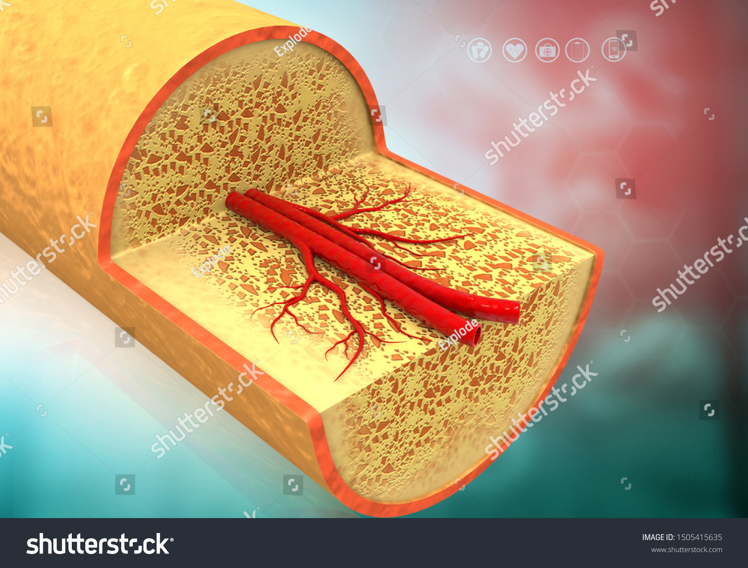
Internal structure of a human long bone, with a magnified cross section of the interior. A cross section of a human long bone. The outlined area is a cross section of an osteon of compact bone. Download this cross section anatomy of human bone photo now. Concentric layers of bone cells (osteocytes) and bone matrix surround the central canal. Find the perfect cross section bone human stock photo. Compact bone cross section courtesy: Select from premium human bone cross section of the highest quality. Dry bone is cut and polished before mounting on a slide. The central tubular region of the bone, called the diaphysis, flares outward near the end to form the metaphysis, which contains a largely cancellous, or spongy, interior. Cross section of a muscular artery showing the smooth muscle in the extensive. Related posts of cross section of a long bone bone test anatomy and physiology. To the left is muscle tissue, and to the right is bone marrow.
No need to register, buy now! The central tubular region of the bone, called the diaphysis, flares outward near the end to form the metaphysis, which contains a largely cancellous, or spongy, interior. In the center of each osteon is the central canal, a space that houses blood vessels and nerves that supply bone. Related posts of cross section of human bone diagram bone in arm pictures. Download this cross section anatomy of human bone photo now.
Cross Section Anatomy Human Bone 3d Stock Illustration 1505415635 from image.shutterstock.com Dry bone is cut and polished before mounting on a slide. Cross sectional anatomy, timothy f. Find the perfect human bone cross section stock photos and editorial news pictures from getty images. The single bone in the thigh is called the femur.this bone is very thick and strong (due to the high proportion of bone tissue), and forms a ball and socket joint at the hip, and a modified hinge joint at the knee. After a fracture, woven bone forms initially and is gradually replaced by lamellar bone during a process known as bony substitution. Human kidney medical model with a cross section of the inner organ with red and blue arteries and adrenal gland as a health care and medical of the anatomy of the urinary system. Compact bone cross section courtesy: In human anatomy, the thigh is the area between the hip and the knee.anatomically, it is part of the lower limb.
No need to register, buy now!
Related posts of cross section of a long bone bone test anatomy and physiology. Compact bone cross section courtesy: At the end of the bone is the epiphysis, which in young people is separated from the. Concentric layers of bone cells (osteocytes) and bone matrix surround the central canal. This is female cross sectional anatomy They are obtained by taking imaginary slices perpendicular to the main axis of organs, vessels, nerves, bones, soft tissue, or even the entire human body. Huge collection, amazing choice, 100+ million high quality, affordable rf and rm images. The inner portion of the bone is composed of trabecular bone and the intervening. A cross section of a human long bone. Bone in arm pictures 12 photos of the bone in arm pictures bone cancer arm pictures, pictures of bone cancer in arm, bone, bone cancer arm pictures, pictures of bone cancer in arm. Internal structure of a human long bone, with a magnified cross section of the interior. Find the perfect cross section bone human stock photo. Select from premium human bone cross section of the highest quality.
The inner portion of the bone is composed of trabecular bone and the intervening. They are obtained by taking imaginary slices perpendicular to the main axis of organs, vessels, nerves, bones, soft tissue, or even the entire human body. Browse 4,294 bone cross section stock photos and images available, or search for human bone cross section to find more great stock photos and pictures. The arrows point toward the tumor. Dutra, human anatomy, anatomical sections, ct scan, computed axial tomography, mri scan, magnetic resonance imaging, virtual autopsy, physician, medical student, reference.
Diagram Of A Rib Cross Section Download Scientific Diagram from www.researchgate.net Cross section of a muscular artery showing the smooth muscle in the extensive. Bone in arm pictures 12 photos of the bone in arm pictures bone cancer arm pictures, pictures of bone cancer in arm, bone, bone cancer arm pictures, pictures of bone cancer in arm. Bone test anatomy and physiology 12 photos of the bone test anatomy and physiology anatomy and physiology bone lab test, anatomy and physiology bone markings test, anatomy and physiology bone practical test, anatomy and physiology bone tissue test, anatomy and physiology test on bone tissue, bone, anatomy and. Concentric layers of bone cells (osteocytes) and bone matrix surround the central canal. Dutra, human anatomy, anatomical sections, ct scan, computed axial tomography, mri scan, magnetic resonance imaging, virtual autopsy, physician, medical student, reference. Last revised on january 1, 2021 The central tubular region of the bone, called the diaphysis, flares outward near the end to form the metaphysis, which contains a largely cancellous, or spongy, interior. The arrows point toward the tumor.
Cross section of a muscular artery showing the smooth muscle in the extensive.
A uniform cross section is the cross section of the solid, parallel to base, such that the resulting figure has the same shape and size as that of the base of the figure.more about uniform cross sectionsolids like pyramids and. Dutra, human anatomy, anatomical sections, ct scan, computed axial tomography, mri scan, magnetic resonance imaging, virtual autopsy, physician, medical student, reference. In three dimensions an osteon is cylindrical in shape. Bone test anatomy and physiology 12 photos of the bone test anatomy and physiology anatomy and physiology bone lab test, anatomy and physiology bone markings test, anatomy and physiology bone practical test, anatomy and physiology bone tissue test, anatomy and physiology test on bone tissue, bone, anatomy and. Compact bone cross section courtesy: The arrows point toward the tumor. Eye anatomy, human eye cross section physiology, cornea model from plastic on wooden table in. This slide contained a cross section of a very small bone, and you are looking at the entire thickness of the shaft of the bone. A cross section of a human long bone. Huge collection, amazing choice, 100+ million high quality, affordable rf and rm images. Smooth muscle and endothelium in a muscular artery wall, (magnification x100). Use scroll bar to go to section of interest you may enlarge any image by 'clicking' on it. They are obtained by taking imaginary slices perpendicular to the main axis of organs, vessels, nerves, bones, soft tissue, or even the entire human body.
Bone in arm pictures 12 photos of the bone in arm pictures bone cancer arm pictures, pictures of bone cancer in arm, bone, bone cancer arm pictures, pictures of bone cancer in arm bone cross section. Internal structure of a human long bone, with a magnified cross section of the interior.


0 Komentar